Breast Cancer Screening for Dense Breasts: What Physicians Should Know
Tanya Gupta, M.D.
Medical Director
Why breast density is more than a mammographic finding in personalized breast cancer screening
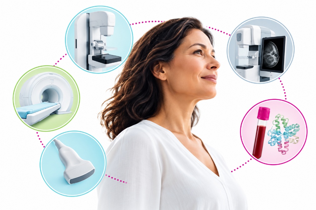
Mammography remains the foundation of breast cancer screening, but its performance varies with breast tissue composition. For women with dense breasts, there are two important implications: dense fibroglandular tissue can obscure malignancies on mammography, and breast density itself is associated with increased cancer risk.
With multiple supplemental screening technologies now available, clinicians have more opportunities to tailor care. Understanding how these approaches differ and where evidence supports their use is essential.
Why Breast Density Changes the Screening Conversation
Breast density is classified using the American College of Radiology's Breast Imaging Reporting and Data System (BI-RADS)1:
-
BI-RADS A: Almost entirely fatty
-
BI-RADS B: Scattered fibroglandular density
-
BI-RADS C: Heterogeneously dense
-
BI-RADS D: Extremely dense
Categories C and D are classified as dense breasts.
The clinical concern is masking: both dense tissue and many breast cancers appear white on mammograms, potentially making abnormalities more difficult to identify.
Since September 2024, FDA regulations have required mammography facilities to notify patients about their breast density.2 For ordering physicians, this creates an important opportunity to discuss individual risk and whether additional screening is appropriate.
How Sensitive Are Breast Cancer Screening Options in Dense Breasts?
The following modalities provide different types of information. Importantly, published sensitivity estimates depend on the population studied along with whether the examination was performed for screening or diagnostic evaluation.
2D Mammography: The Established Foundation
Digital mammography remains an essential screening tool with demonstrated mortality benefits. However, sensitivity decreases as breast density increases, particularly in women with extremely dense breasts.
For patients with BI-RADS C or D density, a negative mammogram does not eliminate the possibility of breast cancer, and mammographic sensitivity is lower in women with dense breasts.
3D Mammography: Improving Detection Through Tomosynthesis
Digital breast tomosynthesis (DBT), commonly called 3D mammography, reduces the effect of overlapping tissue by reconstructing breast images into thin sections.
In a systematic review and meta-analysis published online in 2024 in Advances in Clinical and Experimental Medicine, Lin and colleagues analyzed 11 studies involving more than 2.1 million individuals and reported pooled sensitivity of 91% for DBT versus 86% for digital mammography.3 However, these estimates included women with both dense and non-dense breasts and were not specific to women with BI-RADS C or D breast density.
Importantly, breast density can substantially affect performance. In a 2020 study published in JAMA, Comstock and colleagues reported DBT sensitivity of just 39.1% among women with dense breasts (BI-RADS C and D), compared with 95.7% for abbreviated breast MRI.4 The study included 1,444 women undergoing screening, with 23 breast cancer cases identified.
These findings reinforce that while DBT improves cancer detection overall, significant masking challenges can remain in women with dense breast tissue, supporting individualized discussions about supplemental screening.
Contrast-Enhanced Mammography: Adding Functional Information
Contrast-enhanced mammography (CEM) combines mammographic imaging with iodinated contrast to visualize areas of increased vascularity associated with malignancy.
CEM has demonstrated high diagnostic sensitivity in women with dense breasts. A 2024 systematic review and meta-analysis by Lin and colleagues, published in Cancer Medicine, evaluated 10 studies involving 827 patients with dense breasts and suspicious findings.5 The investigators reported 95% sensitivity (95% CI, 92%–97%) and 81% specificity (95% CI, 70%–89%). These findings reflect diagnostic evaluation rather than screening of asymptomatic women.
Evidence also supports CEM's potential role in supplemental screening. In the 2025 BRAID randomized controlled trial, published in The Lancet, supplemental CEM detected 19.2 additional cancers per 1,000 examinations among women with dense breasts (BI-RADS C and D) following negative mammography.6
Together, these findings suggest that CEM can provide valuable additional information when dense tissue limits conventional mammographic detection. However, iodinated contrast exposure, access, and patient-specific contraindications must be considered when selecting supplemental screening.
Ultrasound: An Accessible Supplemental Option
In some instances, breast ultrasound can detect mammographically occult cancers.
Its advantages include broad availability and utility for targeted evaluation. However, performance varies with examination technique and patient population, and supplemental ultrasound can increase recalls and benign biopsies.
In a 2024 study published in Radiology, Sprague and colleagues evaluated 34,791 supplemental ultrasound screening examinations in women with dense breasts.7 Ultrasound detected 2.0 cancers per 1,000 examinations overall, increasing to 5.5 per 1,000 among women at higher risk of advanced breast cancer. However, the study also reported 29.6 false-positive biopsy recommendations per 1,000 examinations.
In the BRAID trial, automated breast ultrasound identified 4.2 additional cancers per 1,000 examinations in women with dense breasts and negative mammograms.6
Ultrasound may be particularly useful when MRI is unavailable or unsuitable, although it generally detects fewer additional cancers than contrast-enhanced MRI.
Breast MRI: High Sensitivity for Selected Patients
Contrast-enhanced breast MRI provides highly sensitive detection and is an important supplemental screening modality for women at elevated breast cancer risk.
A 2025 meta-analysis by Lian and colleagues evaluated MRI combined with mammography in women with dense breasts and reported 87% pooled sensitivity and 95% specificity for the combined approach.8
Additional evidence demonstrates MRI's ability to detect cancers that may be missed by mammography in dense breast tissue. In a 2020 study published in JAMA, Comstock and colleagues reported 95.7% sensitivity for abbreviated breast MRI compared with 39.1% for digital breast tomosynthesis (DBT) among women with dense breasts (BI-RADS C and D).4 These results highlight the potential value of MRI in patients for whom dense tissue limits mammographic detection.
More recently, the 2025 BRAID randomized controlled trial reported 17.4 additional cancers detected per 1,000 abbreviated MRI examinations following negative mammography in women with dense breasts.6
MRI's clinical advantages must be balanced against cost, availability, intravenous gadolinium contrast, examination tolerance, and the potential for false-positive findings and additional biopsies.
Certitude®: A Proteomic Approach to Breast Cancer Detection
Unlike imaging technologies, Certitude is a blood-based test that uses AI-driven deep proteomic analysis to identify protein patterns associated with breast cancer. Rather than visualizing breast anatomy, the test evaluates circulating biological signals, offering a different approach to breast cancer detection.
In a study published in Endocrinology in 2026, Horrmann and colleagues evaluated 335 women, including 116 patients with newly diagnosed Stage 0–II breast cancer and 219 healthy controls.9 The proteomic classifier demonstrated 86.2% sensitivity (95% CI, 78.8-91.3%) and 90.4% specificity (95% CI, 85.8-93.6%). Importantly, when density information was reported, sensitivity remained high across breast density categories, reaching 90.2% in women with dense breasts (BI-RADS C/D) and 87.2% in women with non-dense breasts (BI-RADS A/B), at 90% specificity.
A second study by Drake and colleagues, published in Breast Cancer: Targets and Therapy in 2026, further evaluated the approach using a larger biobanked sample collection. In a held-out validation cohort of 397 women, the classifier demonstrated 92.6% sensitivity and 92.3% specificity, including 88.6% sensitivity for Stage I and 96.9% for Stage II breast cancer.10
These findings support the potential of deep proteomic testing as an additional source of information in breast cancer detection. However, the studies used case-control and biobanked samples rather than prospective supplemental screening populations. The larger validation study did not record breast density, and its reported 99.87% negative predictive value was modeled using an assumed breast cancer prevalence of 1.25%, rather than measured directly in a prospective screening cohort.
Although prospective screening studies are underway to establish its clinical utility, the published evidence provides a foundation for physicians to evaluate this approach today.
What Do Current Screening Guidelines Recommend?
Recommendations differ in how they interpret the evidence for supplemental screening.
The American College of Radiology's 2024 Appropriateness Criteria update, published in 2025, supports risk- and density-based selection of supplemental modalities.11 Breast MRI or abbreviated MRI is usually appropriate for women at high risk, regardless of density. MRI, ultrasound, and, in selected circumstances, CEM may be considered for other risk-density combinations.
The 2024 U.S. Preventive Services Task Force recommends biennial mammography for women ages 40–74 but concludes that evidence remains insufficient to assess the balance of benefits and harms of supplemental MRI or ultrasound solely based on dense breasts after a negative mammogram.12
European guidance supports offering supplemental MRI to women with extremely dense breasts, emphasizing shared decision-making.
These differences do not eliminate the need for individualized care. They reinforce the importance of considering both the available evidence and the patient's overall risk.
Helping Patients Navigate Screening Decisions
For a busy OB-GYN or primary care physician, a practical approach begins with three questions:
-
What is the patient's breast density and overall risk? Consider family history, genetic pathogenic variants, prior breast lesions, previous chest irradiation, and validated risk assessment.
-
What clinical question remains after mammography? Supplemental screening after a negative examination is different from diagnostic evaluation of a suspicious finding.
-
Which evidence-supported option is accessible and appropriate? Consider detection performance, false-positive burden, contraindications, patient preferences, and the follow-up pathway.
A negative supplemental test should not delay indicated diagnostic imaging or biopsy when clinical or imaging findings remain concerning.
More Information. More Individualized Care.
The future of breast cancer screening is not necessarily one technology replacing another. It is the thoughtful integration of complementary information.
Learn more about Astrin's research into Certitude and its ongoing evaluation as an emerging approach to breast cancer detection.
Footnotes
-
FDA Updates Mammography Regulations to Require Reporting of Breast Density Information and Enhance Facility Oversight | FDA ↩
-
Diagnostic accuracy of digital breast tomosynthesis and digital mammography in women with dense or non-dense breast tissue: A systematic review and meta-analysis - PubMed ↩
-
Comparison of Abbreviated Breast MRI vs Digital Breast Tomosynthesis for Breast Cancer Detection Among Women With Dense Breasts Undergoing Screening | Radiology | JAMA | JAMA Network ↩ ↩2
-
Diagnostic performance of contrast-enhanced mammography for suspicious findings in dense breasts: A systematic review and meta-analysis - PubMed ↩
-
Comparison of supplemental breast cancer imaging techniques—interim results from the BRAID randomised controlled trial - The Lancet ↩ ↩2 ↩3
-
Performance of Supplemental US Screening in Women with Dense Breasts and Varying Breast Cancer Risk: Results from the Breast Cancer Surveillance Consortium - PMC ↩
-
Meta-analysis of the synergistic effect of magnetic resonance imaging and mammography in breast cancer detection in women with dense breasts - PubMed ↩
-
Plasma-based Deep Proteomic Platform for early-stage Breast Cancer Detection | Endocrinology | Oxford Academic ↩
-
Development and Validation of a Machine-Learning Deep Plasma Proteome | BCTT | Dove Medical Press ↩
-
ACR Appropriateness Criteria® Imaging of Invasive Breast Cancer - Journal of the American College of Radiology ↩
-
Recommendation: Breast Cancer: Screening | United States Preventive Services Taskforce ↩