What Every Physician Needs to Know About Dense Breasts
Badrinath Konety, M.D.
Chief Medical Officer
Understanding Breast Density and Its Impact on Screening Decisions
Breast density has become an increasingly important consideration in breast cancer screening. As breast density notification laws and updated FDA requirements have expanded patient awareness, more women are asking, "What does it mean to have dense breasts, and what should I be doing differently?"
For providers caring for women with dense breasts, understanding how density influences both cancer risk and imaging performance is essential. Today's screening landscape offers more options than ever including mammography, breast MRI, ultrasound, and emerging blood-based tests, but each plays a different role within comprehensive breast care.
What Are Dense Breasts?
Breast density describes the proportion of fibroglandular tissue compared with fatty tissue seen on a mammogram. It is categorized using the BI-RADS® breast density classification1:
- A: Almost entirely fatty
- B: Scattered fibroglandular density
- C: Heterogeneously dense
- D: Extremely dense
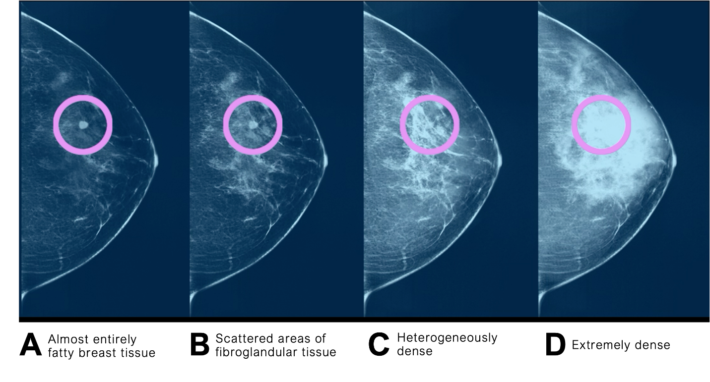
Women classified as BI-RADS C or D are considered to have dense breasts.
Dense breast tissue is common. Approximately 40–50% of women undergoing screening mammography have dense breast tissue, making it one of the most frequently encountered findings in breast imaging.2
Importantly, breast density is not something patients or physicians can feel during a physical exam. It can only be determined through mammographic imaging.
What Do BI-RADS Breast Density Categories Mean?
The American College of Radiology's Breast Imaging Reporting and Data System (BI-RADS®) classifies breast composition into four categories, A through D.3 These categories help physicians understand how breast tissue composition may affect mammographic sensitivity and inform individualized screening discussions.
BI-RADS A: Almost Entirely Fatty
Breasts contain predominantly fatty tissue with minimal fibroglandular density. Mammographic sensitivity is generally highest in this category because there is less dense tissue to obscure potential abnormalities.
Clinical consideration: Continue guideline-recommended mammography, with additional evaluation determined by overall risk, symptoms, or imaging findings rather than density alone.
BI-RADS B: Scattered Areas of Fibroglandular Density
Breasts contain scattered areas of dense glandular and fibrous tissue, but most breast tissue remains non-dense.
Importantly, BI-RADS B is not classified as dense breast tissue. Only categories C and D meet the mammographic definition of dense breasts.
Clinical consideration: Routine screening remains appropriate for average-risk patients. A BI-RADS B classification does not independently indicate a need for supplemental screening, although other risk factors or clinical findings may warrant additional evaluation.
BI-RADS C: Heterogeneously Dense
Substantial areas of fibroglandular tissue are present and may obscure small masses on mammography. BI-RADS C is classified as dense breast tissue.
Clinical consideration: Incorporate breast density into the patient's overall breast cancer risk assessment. Discuss the potential limitations of mammography and whether supplemental screening is appropriate based on lifetime risk, medical history, and shared decision-making.
BI-RADS D: Extremely Dense
Breasts contain extensive fibroglandular tissue, which can substantially reduce mammographic sensitivity. BI-RADS D is the highest density category and is associated with increased breast cancer risk compared with predominantly fatty breasts.
Clinical consideration: Review the patient's comprehensive risk profile and consider whether additional screening evaluation is appropriate. Extremely dense tissue warrants particular attention to mammographic masking, but supplemental screening decisions should still reflect individualized risk and current clinical guidance.
Why Breast Density Matters
Dense breasts matter for two primary reasons.
1. Increased Breast Cancer Risk
Dense breast tissue is an independent risk factor for breast cancer. Women with extremely dense breasts have a significantly higher risk of developing breast cancer than women with predominantly fatty breast tissue.4
Breast density should therefore be considered alongside other established risk factors, including:
- Family history
- Genetic mutations (such as BRCA1/2)
- Prior atypical breast lesions
- Previous chest radiation
- Personal history of breast cancer
- Reproductive history and hormonal exposures
While breast density alone does not necessarily place a patient into a high-risk category, it should be incorporated into individualized risk assessment models whenever appropriate.
2. Reduced Mammographic Sensitivity
Dense fibroglandular tissue appears white on a mammogram—the same color as many breast cancers. This "masking effect" can make small cancers more difficult to detect, reducing the sensitivity of mammography in women with dense breasts.
This is one reason why interval cancers—those diagnosed between routine screening examinations—occur more frequently among women with dense breast tissue.5
Translating Breast Density into Clinical Decisions
Breast density categories describe tissue composition, not the likelihood that a specific mammographic finding is malignant. They should not be confused with BI-RADS assessment categories, which communicate imaging findings and recommended management.
For physicians, the next step is to integrate breast density with established risk factors, mammographic findings, and patient preferences. BI-RADS C and D warrant a discussion about breast density and its implications, but density classification alone does not determine the appropriate screening or diagnostic pathway.
This distinction supports personalized breast care without replacing established screening recommendations.
Talking to Patients About Dense Breasts
Patients often interpret a dense breast notification as alarming. Clear communication can help reduce unnecessary anxiety while reinforcing the importance of continued screening.
Helpful discussion points include:
- Dense breasts are common.
- Dense breast tissue does not mean cancer is present.
- Mammography remains essential.
- Additional screening may be appropriate based on overall risk—not density alone.
- Screening recommendations should be individualized.
Shared decision-making remains central to selecting supplemental screening strategies.
What Current Guidelines Recommend
Major organizations including the American College of Radiology (ACR), American College of Obstetricians and Gynecologists (ACOG), and American Cancer Society (ACS) agree on several key principles:
- Mammography remains the primary screening test.
- Breast density should be included in individualized risk assessments.
- Women at elevated lifetime risk may benefit from supplemental MRI.
- Decisions regarding supplemental screening should consider breast density, overall risk, patient preferences, and access to care.
There is no one-size-fits-all approach for women with dense breasts.
Bringing Greater Confidence to Dense Breast Care
As breast cancer screening evolves, physicians have more opportunities to personalize care than ever before.
Understanding breast density is the first step. Combining individualized risk assessment with appropriate use of mammography, MRI, ultrasound, and emerging blood-based testing allows clinicians to build screening strategies tailored to each patient.
Certitude is designed to provide another layer of insight for women with dense breasts when standard screening leaves questions—supporting informed clinical decisions while fitting seamlessly into existing workflows.
Interested in learning how Certitude may complement your breast screening pathway? Learn more about integrating Certitude into your practice and discover how a blood-based option can support confident, patient-centered care.